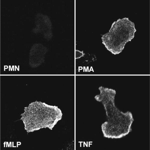
Fig. 3.

Detecting upregulation of proteins at the cell surface. Neutrophils plated on fibrinogen-coated coverslips were left untreated or stimulated with 200 nM PMA, 1 μM fMLF, or 0.02 μM TNFα as indicated. Fixed, intact cells were stained with mAb 7D5 to show upregulation of flavocytochrome b558 at the cell surface. All four images were acquired using identical confocal settings
