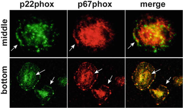
Fig. 5.

Localization of NADPH oxidase subunits. Neutrophils were plated on fibrinogen-coated coverslips and then stimulated with TNFα for 30 min. Fixed and permeabilized cells were double stained to detect p22phox (green) and p67phox (red). Confocal sections taken through the center of cells or at the substrate-adherent surface indicate colocalization of NADPH oxidase subunits at the leading edge and throughout the plasma membrane (arrows). In contrast, p67phox was not detected on p22phox -positive specific granules (arrowheads)
