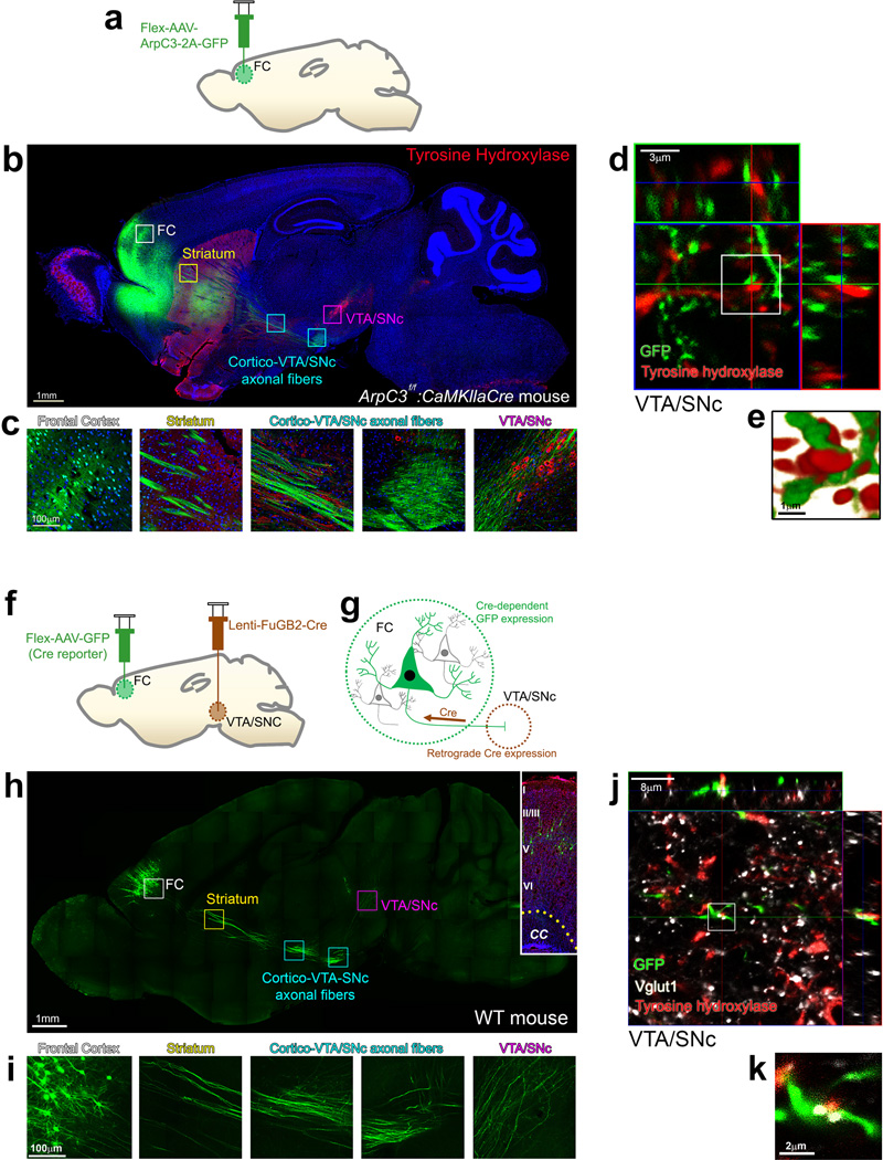
Fig. 3. Arp2/3 rescued excitatory neurons of the frontal cortex project to and make synaptic contacts within the VTA/SNc.
(a) Schematic representation of the rescue virus (Flex-AAV-ArpC3-2A-GFP) injection into the frontal cortex (FC). (b) Representative sagittal section image of GFP (green) expression and immunostaining for tyrosine hydroxylase (red) from an Arp2/3 frontal cortical rescue mouse. Boxes represent higher magnification images in (c). (c) High magnification images tracing the GFP positive neurons and their afferents from the FC all the way to the ventral tegmental area (VTA)/substantia nigra (SNc). (d) Representative maximum projection image with orthogonal views of GFP positive axons (green) and tyrosine hydroxylase immunohistochemistry (red) within the VTA/SNc. GFP within axons is from an ArpC3f/f: CaMKIIαCre mouse with Flex-AAV-ArpC3-2A-GFP virus injected into the FC. (e) High magnification surface rendering depicting contact between FC axons and tyrosine hydroxylase positive neurons within the VTA/SNc. (f and g) Schematic representation of the retrograde viral tracing between the VTA/SNc and FC. (h) Representative sagittal section visualizing Cre-dependent GFP expression in the FC mediated by a Cre-expressing rabies/lenti-viral injection (Lenti-FuGB2-Cre) into the VTA/SNc. Boxes represent higher magnification images in (i). Inset shows GFP-positive neurons from a FC section stained with DAPI (blue) and NeuroTrace® (red) to visualize the cortical layers. CC, corpus callosum. (i) High magnification images tracing the GFP positive neurons and their afferents from the FC all the way to the VTA/SNc. (j) Representative maximum projection image of GFP positive axons (green) labeled by retrograde lenti-FuGB2-Cre tracing from the VTA/SNc. Vglut1 and tyrosine hydroxylase immunohistochemistry labels dopamine-producing neurons (red) and presynaptic terminals (white). (k) High magnification view of co-localized axons (green), excitatory presynaptic marker (white) and dopamine neurons (red) within the VTA/SNc. All representative images were successfully repeated more than three times.

