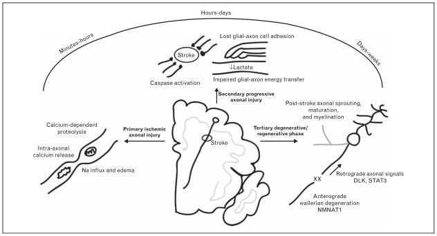
FIGURE 3.
Schematic figure illustrating the three distinct phases of axonal injury and repair after stroke. As an example, focal lacunar stroke within the centrum semiovale is represented. The primary ischemic phase results from catastrophic failure of adenosine triphosphate availability, leading to Na+ retention, axonal swelling, and intra-axonal Ca2+ release from axoplasmic reticulum. These events trigger calcium-dependent proteolysis and potentially activate the secondary progressive phase of injury. Multiple molecular targets participate in this phase including caspase activation, loss of axoglial contact and/or failure of glial-to-axon energy transfer. A third phase begins in the days to weeks after stroke and involves a combination of anterograde Wallerian degeneration, retrograde axonal injury signaling and activation of axonal sprouting or degenerative pathways.

