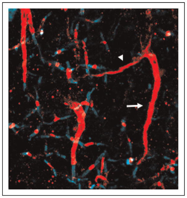
FIGURE 4.
Identification of supranumerary axon initial segments in peri-infarct cortex. Immunofluorescent labeling of the axon initial segment (beta-IV spectrin, red) allows identification of a typical axon initial segment projecting towards white matter (arrow). Arising laterally and projecting intracortically is a thin, shorter axonal initial segment (arrowhead). Labeling for contactin-associated protein (caspr, teal) shows the distal end of the original axon initial segment is myelinated while the newly sprouted axonal initial segment remains unmyelinated at its distal end.

