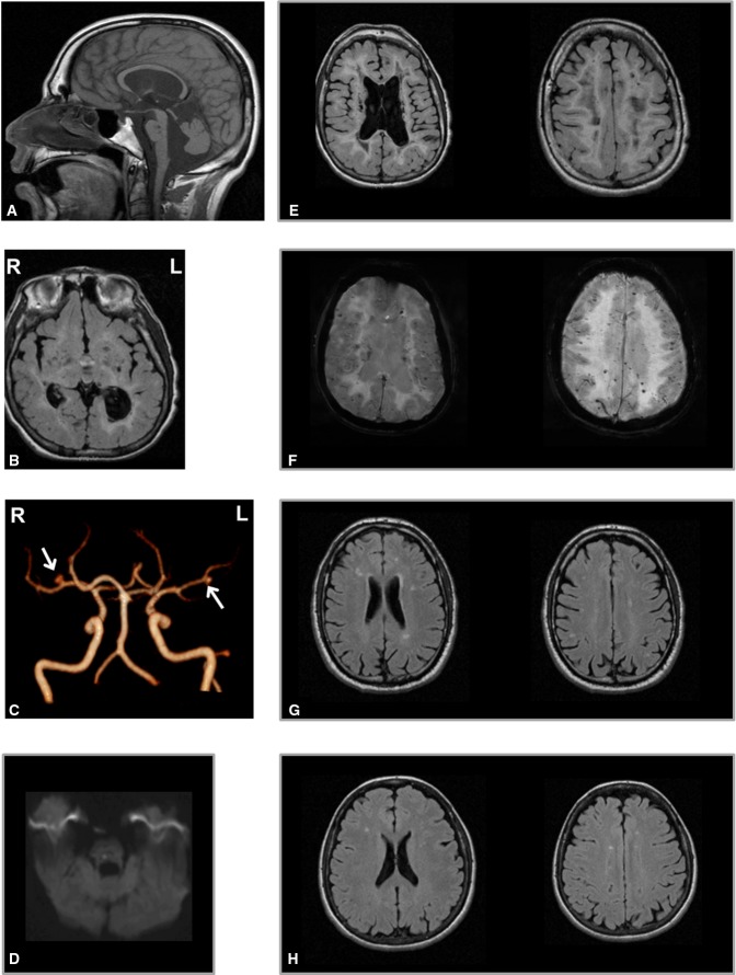
Figure 1.
- A–F Brain MRI of the proband at 23 years. (A) Sagittal fast spin echo (FSE) T1-weighted image shows thinning of the corpus callosum, dilation of the IV ventricle and of the cisterna magna and reduced volume of vermis and brainstem; two lacunar lesions are evident in the dorsal pons. (B) T2-weighted axial fluid-attenuated inversion recovery (FLAIR) images show hyperintense periventricular white matter with several lacunar lesions also in the basal ganglia and thalami. (C) MR angiography 3D TOF (time of flight) reconstruction shows two saccular aneurisms in the right M1 (ø 4 mm) and left M2 (ø 2.5 mm) segments of middle cerebral arteries (arrows). (D) A recent ischemic hyperintense lesion is detected on diffusion tensor imaging (DTI) in the right side of the dorsal pons. Severe cavitations and lateral ventricles dilation (left > right) on FLAIR images (B, E) and diffuse microbleeds as small hypointense foci on SWI are visible (F) in the same slices of (E). R = right, L = left.
- G, H Brain MRI scans of the asymptomatic parents of the proband show multiple focal hyperintensities on T2-weighted images in the periventricular and subcortical cerebral white matter, expression of gliosis secondary to chronic small vessel ischemic changes, more evident in the father (G), 56 years, than in the mother (H), 54 years.

