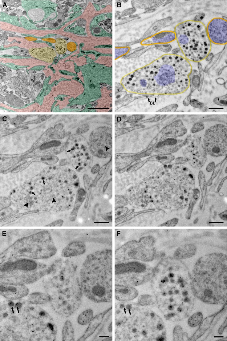
Fig 4. Serial sections show subcellular distribution of vGluT2.
Images were taken from a representative middle-aged vehicle treated female rat. (A) A low magnification electron microscopy image was pseudocolored to show neuroterminals with (light yellow) and without (orange) large dense-core vesicles, a convoluted basal lamina covered portal capillary area—typical in older rats (pink), and tanycytic elements (green). (B) A higher magnification image of panel A was shown. This section was used as a negative control, in which omitting primary vGluT2 antibody resulted in no immunogold labels in the terminal. Two neurosecretory terminals containing large dense-core vesicles in direct contact with the basal lamina (BL with arrow) of the portal capillary region were highlighted in light yellow, and surrounding neuronal elements without large secretory vesicles were highlighted in orange. Groups of small vesicles in neurosecretory terminals and surrounding neuronal elements were pseudocolored in blue. A cluster of small vesicles was distributed in the central portion of the lower left terminal. Another cluster of irregular shaped small vesicles was shown to the left. (C, D) Serial sections adjacent to panel B were used to label vGluT2 protein. Immunogold labels for vGluT2 were seen on large dense-core vesicles (arrow). Whether small vesicles (arrowhead) contained vGluT2 immunosignal was difficult to determine, even at higher magnification. (E, F) By examining adjacent sections, it could be seen that immunogold labels of vGluT2 were present in most dense-core vesicles. As indicated by double arrows, some large dense-core vesicles in image E showed no immunogold labels, while the same vesicle in the adjacent image F was immunolabeled. This is likely to be the case because the antibody reaction site on large dense-core vesicle (about 120 nm diameter) might only be captured in sections (each about 50 nm thick) in which antigen is exposed. BL (panel B) = basal lamina. Scale bar panel A = 2 μm, B-D = 500 nm, E-F = 200 nm.

