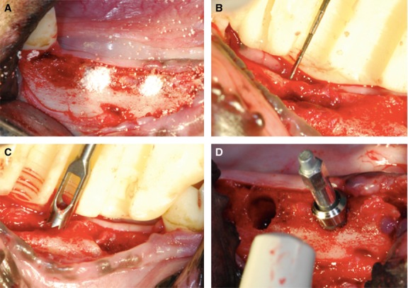
Figure 1.

Cylindrical-defect osteotomy and 4 weeks re-entry surgeries were applied for clinical and histological analysis. (A) Standardized sockets were prepared in both sides of the mandible. The socket defects were divided into three local treatment modalities. (B) The amount of vertical ridge resorption was evaluated according to a surgical stent. (C) The 3-mm-diameter and 6-mm-deep bone cores were drawn with a 4-mm-diameter trephine bur from the sites determined by a surgical stent. (D) A 4.8-mm-diameter with 6-mm-length implant was inserted and the ISQ was detected.
