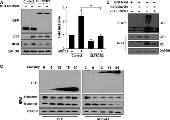
Figure 3.

GLTSCR2 enhances NPM degradation through proteasomal ubiquitination pathway. (A) HeLa cells were transfected with pGFP (control) or pGFP-GLT (GLTSCR2) for 48 hrs. Cells were either not treated or treated with 20 μM MG132 for 6 hrs and cell lysates were Western blotted using the indicated antibodies (left panel). The histogram plot shown is the densitometric quantification of NPM after normalization to MG132-untreated pGFP-transfected cells (mean ± SD; *P < 0.05). (B) HeLa cells were transfected with the indicated plasmids. Cells were further treated with 20 μM MG132 for 6 hrs and lysates were pulled down by Ni2+-chelating Sepharose for 3 hrs. The resultant pellets were Western blotted using anti-GFP antibody (upper panel). The expression levels of GFP-NPM, V5-GLTSCR2 and GAPDH are shown in the three lower panels. (C) GLTSCR2 suppresses oligodimerization of NPM. HeLa cells were transfected with pGFP or pGFP-GLT for 24 hrs. Then, cells were treated with 0.1% glutaraldehyde and lysates were Western blotted to detect monomeric or oligomeric forms of NPM after normalization with GAPDH.
