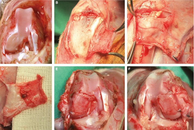
Figure 1.
Surgical technique. (A) Left leg after creation of a critical size cartilage defect of the medial and lateral condyle. (B) Cautious separation of the periosteum flap from the bony surface. (C) Harvesting of the periosteum flap based on the saphenous artery and its venae comitantes, the nourishing vessel to the periosteum can be seen clearly. (D) Bottom surface of the raised flaps; the cambium layer is visible. (E and F) Right and left leg after coverage of the medial defects and fixation with transosseous sutures.

