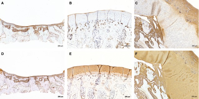
Figure 5.

IHC staining with Coll I (upper row, A–C) and Coll II (lower row, D–F) of corresponding sections of negative control (A and D), positive control (B and E) and condyle covered with periosteum flap (C and F). Coll I staining is found mainly in fibrous tissue (e.g. of the periosteum flap) and bony surface (e.g. of the free subchondral bone in negative control), but not in cartilage, as typical. The original cartilage of the positive control as well as the neo-cartilage show characteristic positive results for Coll II.
