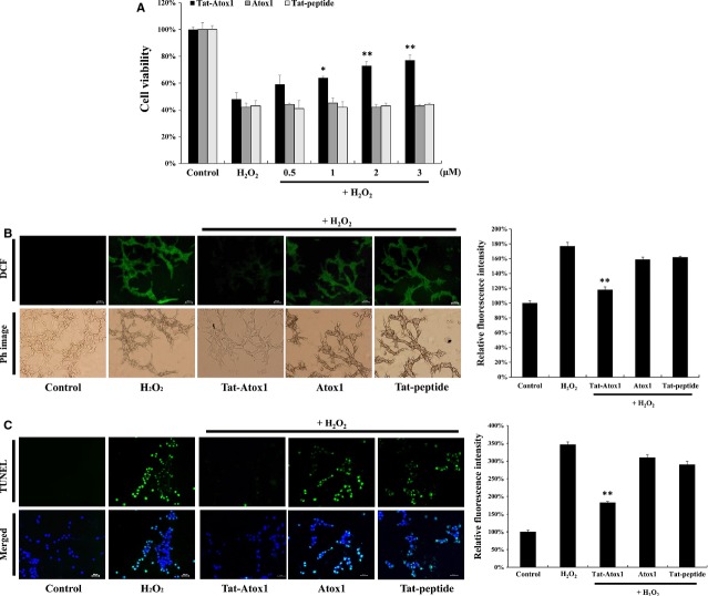
Figure 2.
Protective effects of transduced Tat-Atox1 protein against oxidative stress. Pre-treatment of HT-22 cells with Tat-Atox1 protein (0.5–3 μM) and control Atox1 protein for 1 hr and treatment with 1 mM hydrogen peroxide (H2O2) for 2 hrs. Then, cell viability was assessed by MTT assay (A). *P < 0.05 and **P < 0.01 compared with H2O2-treated cells. Effects of transduced Tat-Atox1 protein on H2O2-induced ROS production. Treatment with Tat-Atox1 protein (3 μM) and control Atox1 protein was followed by 10 min. treatment with H2O2 (1 mM). Intracellular ROS levels were measured by DCF-DA staining and fluorescence intensity was measured by ELISA plate reader (B); scale bar = 50 μm. **P < 0.01 compared with H2O2-treated cells. Effects of transduced Tat-Atox1 protein on H2O2-induced DNA fragmentation. One-hour pre-treatment of HT-22 cells with Tat-Atox1 protein (3 μM) and control Atox1 protein was followed with 4-hr treatment with H2O2 (1 mM). DNA fragmentation was measured by TUNEL staining and the fluorescent intensity was measured by ELISA plate reader (C); scale bar = 50 μm. **P < 0.01 compared with H2O2-treated cells.

