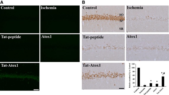
Figure 5.

Effect of transduced of Tat-Atox1 protein on animal brain. Transduction of Tat-Atox1 protein into brain (A). Animals were treated with single i.p. injection of Tat-Atox1 protein and killed after 6 hrs. Transduction of Tat-Atox1 protein into the CA1 region was determined by immunohistochemistry with a rabbit anti-polyhistidine antibody and FITC-conjugated anti-rabbit IgG. Immunohistochemistry for NeuN in the hippocampal CA1 region (B). Control, ischaemia, Tat peptide, Atox1 protein and Tat-Atox1 protein-treated groups 4 days after ischaemia-reperfusion. SP, stratum pyramidale; SO, stratum oriens; SR, stratum radiatum; bar = 50 μm. The relative number of NeuN-immunoreactive neurons versus control group per section in all the groups (n = 5 per group; *P < 0.05, significantly different from the control group, #P < 0.05, significantly different from the ischaemia group). The bars indicate standard error of mean (SEM).
