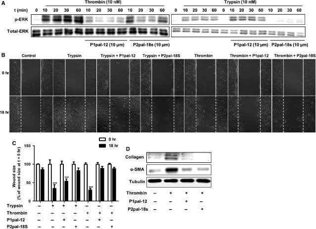
Figure 5.
PAR-2 is required for PAR-1-induced pro-fibrotic responses. (A) Western blot analysis of ERK1/2 phosphorylation in NIH3T3 cells after stimulation with trypsin (10 nM) or thrombin (10 nM), in the absence (-) or presence (+) of P1pal-12 (10 μM) or P2pal-18s (10 μM). P1pal-12 or P2pal-18 sec. was added 30 min. before the stimulation. Total ERK served as loading control. (B) Wound size of NIH3T3 fibroblast monolayers after treatment with DMSO (control), trypsin (10 nM) or thrombin (10 nM) for 18 hrs in the presence or absence of P1pal-12 or P2pal-18s. Cells were pre-incubated with 10 μM P1pal-12 or P2pal-18s for 30 min. as indicated. Shown are photographs of representative microscopic fields. (C) Quantification of the results depicted in (B) as described in the Materials and methods section. Data are expressed as mean ± SEM (n = 6). ***P < 0.001. (D) Western blot analysis of α-SMA and collagen expression in NIH3T3 cells 24 hrs after stimulation with DMSO (control) or thrombin (10 nM), in the presence (+) or absence (-) of P1pal-12 (10 μM) or P2pal-18s (10 μM). Tubulin served as a loading control.

