
Figure 5.
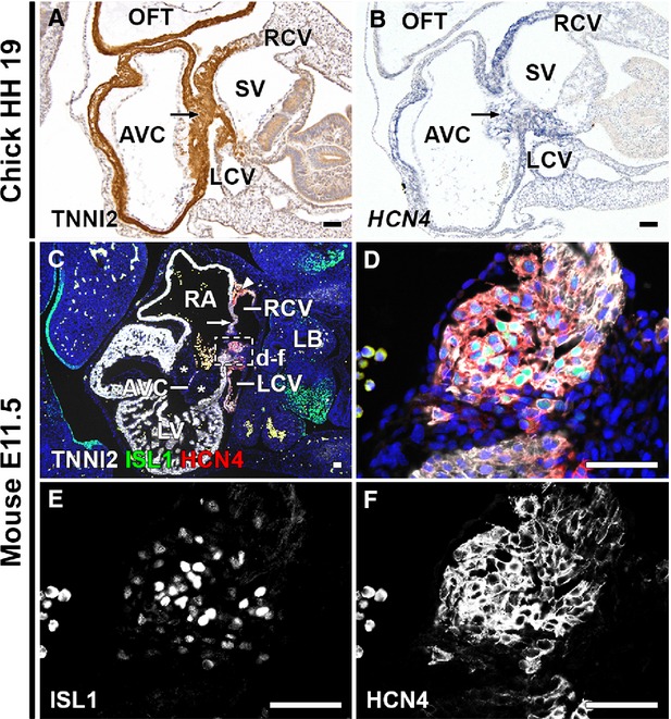
HCN4 expression in myocardial continuity between sinus venosus and posterior AV canal. (A) TNNI2 stain of HH19 chick embryo. Arrow indicates continuity between sinus venosus myocardium and posterior AV canal. (B) ISH shows HCN4mRNA expression in continuity (arrow). (C) Merge of ISL1, TNNI2, HCN4 and DAPI staining of E11.5 mouse embryo, showing the myocardial continuity between the sinus venosus and posterior AV canal. The ISL1+/HCN4+/TNNI2+ SAN (arrowhead) and RVV (arrow) are shown. Note the thick endocardial cushions (asterisks) in the AVC. (D–F) Higher magnifications of boxed area in C and D. ISL1+/TNNI2+/HCN4+ cells are seen in the continuity between sinus venosus myocardium and posterior AV canal myocardium. (E and F) Grey values of ISL1 (E) and HCN4 (F). AVC: atrioventricular canal; LA: left atrium; LB: long bud; LCV: left cardinal vein; LV: left ventricle; OFT: outflow tract; RA: right atrium; RCV: right cardinal vein; SV: sinus venosus; V: ventricle. (C–F) White: TNNI2, green: ISL1, red: HCN4, blue: DAPI; scale bars: 50 μm.