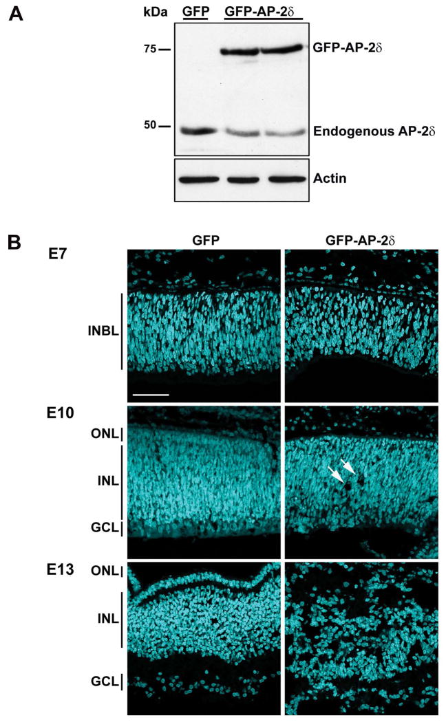
Figure 1. Disruption of the layers of the retina upon overexpression of AP-2δ.
(A) Protein lysates from E9 embryos in ovo electroporated with either GFP or GFP-AP-2δ RCAS expression constructs (two different eyes for GFP-AP-2δ) were separated in a 10% SDS-PAGE gel and transferred to a nitrocellulose membrane. AP-2δ was detected with rabbit anti-AP-2δ antibody. (B) Retinal tissue sections from E7, E10 and E13 embryos in ovo electroporated with GFP or GFP-AP-2δ RCAS expression constructs were stained with DAPI to label the nuclei. INBL, inner neuroblastic layer; ONL, outer nuclear layer; INL, inner nuclear layer; GCL, ganglion cell layer. Photographs were taken with a 40X lens using a Zeiss LSM710 confocal microscope. Arrows indicate holes. Scale bar = 50 μm.

