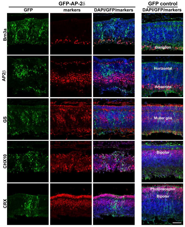
Figure 2. Co-expression of lineage-specific markers and AP-2δ in E10 embryos.
Retinal tissue expressing either GFP or GFP-AP-2δ was double-stained with anti-GFP and anti-Brn3a, anti-AP-2β, anti-GS, anti-CHX10 or anti-CRX antibodies, followed by secondary antibodies linked to Alexa 488 or 555. Sections were counterstained with DAPI to label the nuclei. The location of specific retinal cell types is indicated in GFP control retinal tissue. Photographs were taken with a Zeiss LSM710 confocal microscope equipped with a 40X lens. Scale bar = 50 μm.

