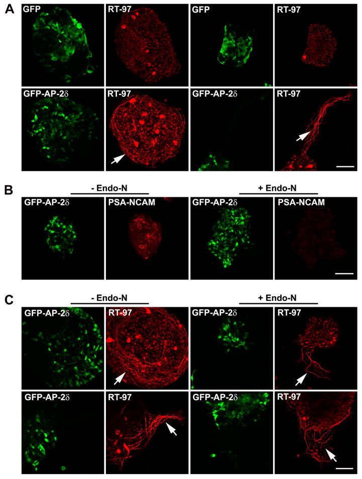
Figure 7. Endo-N treatment relaxes the coils of fibers produced by GFP-AP-2δ-positive cells in vitro.
(A) E2 embryos were in ovo electroporated with GFP or GFP-AP-2δ expression constructs. E7 retinas were dissociated with trypsin and cultured for 48 h. Cells were immunostained with anti-GFP (green) and RT-97 (red) antibodies. Cultures from two separate experiments are shown. Scale bar = 50 μm. (B, C) E2 embryos were in ovo electroporated with GFP-AP-2δ expression construct. E7 retinas were dissociated with trypsin and cultured for 48 h in the presence of Endo-N. Cells were immunostained with anti-GFP (green) and anti-PSA-NCAM (red) (B) or RT-97 (red) (C) antibodies. Cultures from two separate experiments are shown in (C). Scale bar = 50 μm. The arrows point to the coils of RT-97-positive fibers.

