
Fig. 2.
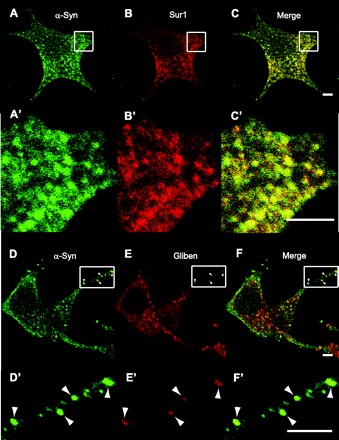
Colocalization of α-synuclein with SUR1 or fluorescent glibenclamide at punctate structures. A: anti-α-synuclein (sc-7011, C20) was used to stain cells isolated from mouse islets and detected by Alexa 488-labeled secondary antibody, as before. A′: magnification of the region indicated by the rectangle in A. B: anti-SUR1 (sc-5789, C16) was used to stain the same cells but detected using secondary antibody conjugated to Alexa fluor 594. B′: magnification of the region indicated by the rectangle in B. C: merge of images in A and B. C′: magnification of the region indicated by the rectangle in C. D–F and D′–F′: same as for A and B and A′ and B′, except that red fluorescence is due to red Bodipy®TR-glibenclamide, rather than SUR1 antibody staining. Shape and pattern of green and red fluorescent puncta show incomplete but clear coincidence patterns, which were typical of all sections through the cell. Scale bars, 2 μm.