


Fig. 3.
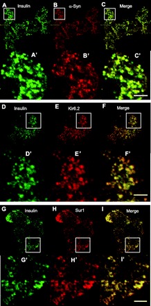
α-Synuclein, Kir6.2, and SUR1 localized to insulin-secretory granules. Anti-insulin (A0564) immunohistochemistry was detected using secondary antibody conjugated to Alexa fluor 488. Anti-α-synuclein (sc-7011, C20), anti-Kir6.2 (sc-11226, N18), and anti-SUR1 (sc-5789, C16) were detected by secondary antibody conjugated to Alexa fluor 594. A–C: anti-insulin (green), anti-α-synuclein (red), and merged staining of isolated islet cells. D–F: anti-insulin (green), anti-Kir6.2 (red), and merged staining of isolated islet cells. G–I: anti-insulin (green), anti-SUR1 (red), and merged staining of isolated islet cells. A′–I′: magnifications of regions indicated by the rectangle in the panel of the corresponding letter. Scale bars, 2 μm.