
Fig. 4.
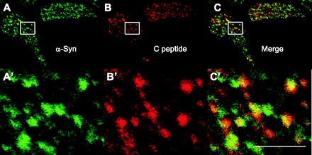
Colocalization of α-synuclein and C-peptide at punctate structures. A: anti-α-synuclein (sc-7011, C20) was used to stain cells isolated from mouse islets and detected by Alexa 488-labeled secondary antibody, as before. B: anti-C-peptide (sc-57046, 5D3) was used to stain the same cells but detected using secondary antibody conjugated to Alexa fluor 594. C: merge of images in A and B. A′–C′: magnifications of regions indicated by the rectangle in the panel of the corresponding letter. In panels labeled by prime letters, scale bar is 2 μm.