
Fig. 9.
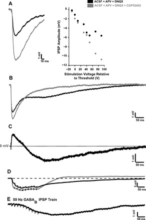
Modulatory effects of GABAB. A: GABAB modulated peak IPSP amplitude. Left, responses of a P28 neuron at (threshold + 90) V stimulation in aCSF + APV + DNQX (black trace) to isolate the IPSP and in aCSF + APV + DNQX + CGP-52432 (gray trace) to block GABAB receptors. Right, responses of the same neuron at varying stimulation amplitudes. Note the increase of IPSP amplitude after GABAB blockade. B: GABAB modulated IPSP temporal properties. Shown is a P17 neuron at (threshold + 50) V stimulation in aCSF + APV + DNQX (black trace) and in ACSF + APV + DNQX + CGP-52432 (gray trace). C: difference waveform of the two traces shown in B. The positive component of the difference waveform corresponds to the increased GABAA IPSP peak amplitude when GABAB receptors are blocked. The negative longer-lasting component of the difference waveform corresponds to the postsynaptic GABAB IPSP that is eliminated with CGP-52432. D: GABAB modulated recovery after repetitive stimulation. P16 neuron responses at 50 Hz in aCSF + APV + DNQX (black trace) and aCSF + APV + DNQX + CGP-52432 (gray trace) are shown. E: GABAB response of a P17 neuron to a 50-Hz train isolated with aCSF + APV + DNQX + picrotoxin.