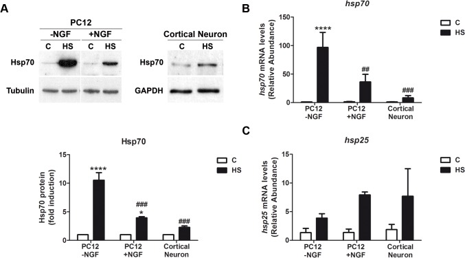
Fig 1. Neuronal cells show weaker induction of Hsp70 in response to heat shock.
(A) Upper panels: Representative immunoblots of Hsp70 protein in undifferentiated (-NGF) and differentiated (+NGF for 7d) PC12 cells, and cortical neurons (E18.5, 7div) subjected to heat shock (42°C, 2h) or kept under control conditions (C, 37°C). Lower panel: Quantification of the relative Hsp70 protein levels showed in the upper panels. Tubulin and GAPDH were used as loading controls. Statistical analyses were performed by Two-way ANOVA followed by Tukey’s multiple comparisons test. ****p < 0.0001, *p < 0.05 compared with control; ###p <0.001, compared to Hsp70 fold induction in undifferentiated PC12 cells during heat shock. (B, C) Relative abundance of hsp70 and hsp25 transcripts in PC12 cells and cortical neurons. hsp70 and hsp25 mRNA levels were calculated comparing the abundance of each cDNA in cells under control and heat shock conditions. For each sample, cyclophilin mRNA was used as a reference gene. Data are expressed as mean plus SEM of at least three independent experiments. Statistical analyses were performed by Two-way ANOVA followed by Tukey's multiple comparisons test. ****p < 0.0001, *p < 0.05 compared with control; ##p < 0.01, ###p <0.001, compared to Hsp70 mRNA abundance in undifferentiated PC12 cells during heat shock.

