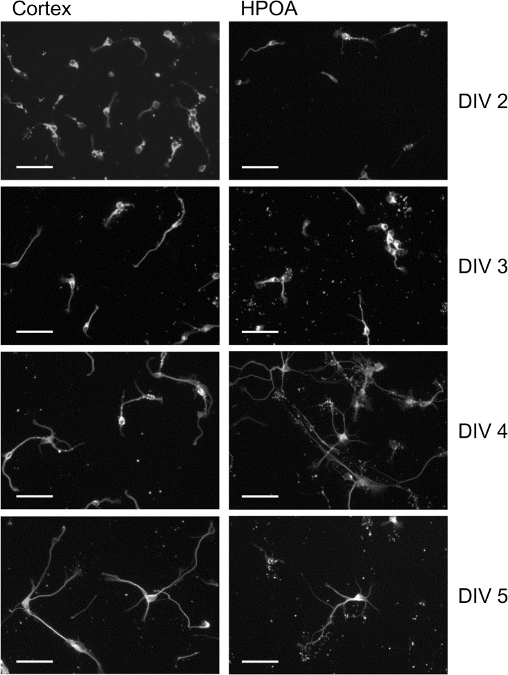
Fig 1. Photomicrographs illustrating the growth of untreated primary neurons derived from GD53 fetal lamb cortex and hypothalamus-preoptic area (HPOA) maintained in vitro for 2 days (DIV2) to 5 days (DIV5).
Soma and neurites were identified by immunohistochemical staining with the neuron-specific mouse anti-β-tubulin antibody. Scale Bar = 50 μm.

