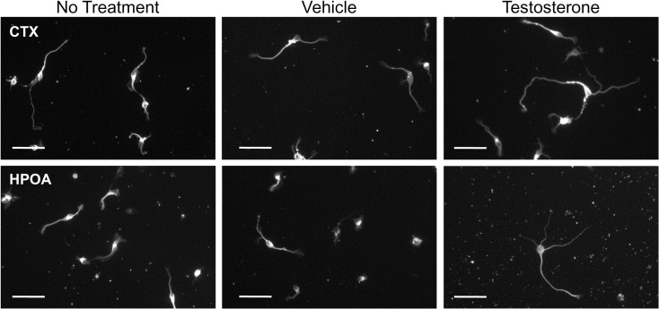
Fig 3. Photomicrographs illustrating the effects of testosterone (10 nM) treatment for 3 days on the morphology of cortical and HPOA neurons immunolabeled for neuron-specific anti-β-tubulin.
Morphometric analysis was carried out on randomly selected neurons from each subgroup. In order to control for possible variations between experimental replicates, untreated cells (No treatment) were evaluated together with control (Vehicle) and testosterone-treated cells. Scale Bar = 50 μm.

