
Fig 5. Extracellular ATP induces NOX2 activation via PKC.
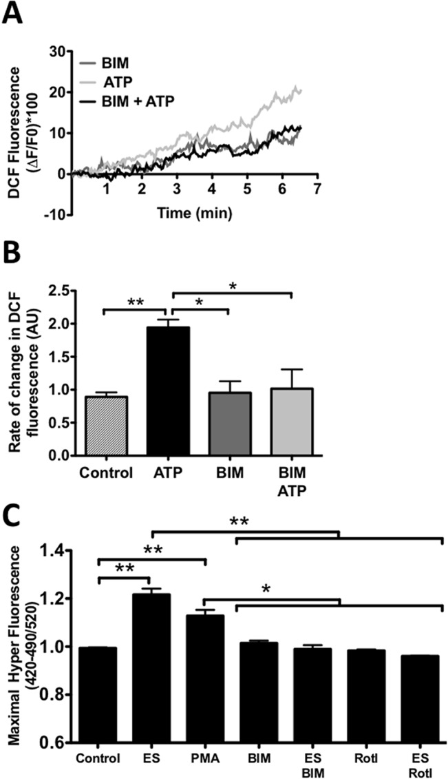
Muscle fibers were isolated, loaded with DCF (30min) and stimulated with exogenous ATP. A, representative traces of DCF fluorescence under control or stimulated with ATP in the absence or presence of BIM (5μM). B, muscle cells were stimulated with ATP and the slope of fluorescence was analyzed (see material and method) (n = 3, *p<0.05). C, muscle fibers were isolated and transfected with HyPer plasmid, 24h post transfection the cells were stimulated in the presence of PMA, BIM or Rotterin (Rotl) as indicated in the graph, maximal fluorescence was plotted (n = 5), *p<0.05, **p<0.01.