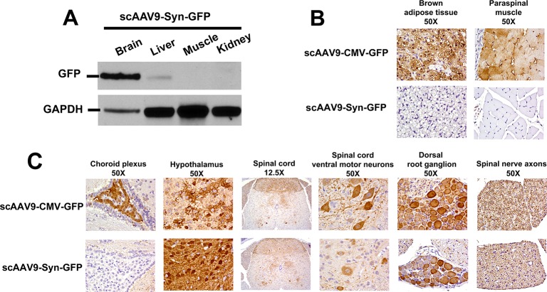
Fig 2. GFP expression driven by the CMV or Syn promoter.
(A) Western blotting analysis of GFP in tissues from animals injected with AAV9-Syn-GFP. GAPDH was used as the loading control. Immunohistochemistry of (B) brown adipose tissue and paraspinal muscle and (C) central nervous system regions. GFP expression (brown) was driven by the CMV or the Syn promoter. AAV particles were injected as described in ‘Material and Methods’ and mice were analyzed 4 weeks thereafter. The results are representative of 2 or more animals. The magnifications are indicated.

