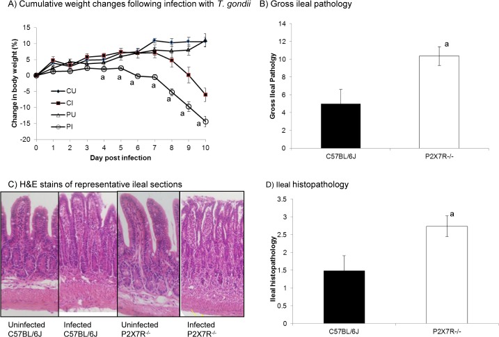
Fig 1. Toxoplasmic ileitis is exacerbated in in P2X7R-/- mice compared with wild type.
Male mice (6–8 weeks old) were infected orally with 10 T. gondii ME49 cysts. (A) Mice were weighed daily for 10 days and cumulative weight changes were calculated and the results presented represent the mean ± SEM of the percentage of weight change relative to starting weight per strain per day from one of six experiments that generated similar data. Infected n = 21/strain; uninfected n = 9/strain. P2X7R-/- mice lost significantly more weight than C57BL/6J mice from day 4 post-infection onward (P<0.005, assessed by multivariate analysis of variance (MANOVA) with days assigned as the within-subjects variable and mouse strain/infection status assigned to the between subjects variable, followed by the assessment of significant interactions within each time point using planned comparisons, i.e., two-way ANOVA coupled to Tukey’s post-hoc test at each day post infection). CU, C57BL/6J uninfected; CI, C57BL/6J infected; PU, P2X7R-/- uninfected; PI, P2X7R-/- infected. (B) Gross ileal pathology was scored on day 8 post-infection based on five observations: consistency of the intestinal contents; absence/presence of blood; absence/presence of pus; degree of swelling; and amount of angiogenesis. This system was adapted from Melgar et al. [60] and was based on an ascending scale of severity, for each parameter, as follows: 0 (no abnormality); 1 (minimal); 2 (moderate); or 3 (severe). The score for each parameter was added to give a total out of a maximum possible score of 15. Gross ileal pathology was also assessed in uninfected mice but no pathology was observed in either strain. Results presented are from one of four experiments that generated similar data. (C, D) Ileal histopathology was evaluated based on five parameters: epithelial cell damage; goblet cell loss; crypt dropout; neutrophil and mononuclear cell infiltration in the submucosa and neutrophil and mononuclear cell infiltration in the muscular layers. Three random fields of view at a 40x magnification were graded on an ascending scale of severity: 0 (no abnormality); 0.25 (minimal); 0.5 (mild); 0.75 (moderate); or 1 (severe) giving a total score out of 5 per mouse. Histopathology was also examined in uninfected mice, however, no pathology was observed in any strain. Photomicrographs of histopathology shown (C) are from single mice and are representative of all mice examined in the group. Results are presented as the mean ± SEM (n = 8) for both strains of mice. Results presented are from one of three experiments that generated similar data. a Indicates where the score is significantly different from the score for C57BL/6J mice (P<0.05, one-way ANOVA coupled to Tukey’s post-hoc test).

