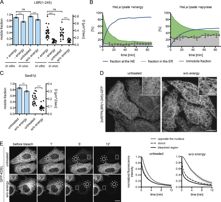
Figure 7.
Energy depletion affects ER structure. (A) Mobile fractions and diffusion coefficient derived from FRAP on 2×RFPtevLBR(1–245)-GFP semipermeabilized reporter cells supplemented with HeLa lysate and either energy or apyrase (in vitro) or intact cells treated with deoxyglucose and NaN3 after 10 min of preincubation (in vivo). Mean ± SEM (error bars); n ≥ 23; ***, P ≤ 0.001 (unpaired t test). (B) Immobile fractions were measured by FRAP in the ER, before (t = 0) and after release of the LBR(1–245) reporter in semipermeabilized cells with HeLa lysate + energy or HeLa lysate + apyrase. INM targeting kinetics were measured as described for Fig. 3 A. The immobile fraction is displayed relative to the whole population. Mean ± SD (error bars). (C) Mobile fractions and diffusion coefficients upon energy depletion derived from FRAP performed on intact cells expressing Sec61β-GFP as in A. Mean ± SEM (error bars); n = 21; **, P ≤ 0.01 (unpaired t test). (D) The ER gets fenestrated upon energy depletion in vivo. Representative images are shown of 2×RFPtevLBR(1–245)-GFP reporter cells treated with deoxyglucose and NaN3 for 20 min before fixation. Maximum intensity projection (1.2 µm) of five confocal slices from the lower cell surface to the NE. (E) FLIP performed on cells transiently transfected with GFP-KDEL, untreated or treated with deoxyglucose and NaN3. Circle, bleached area; dashed circle, “donut” area; square, “opposite the nucleus” area. Mean ± SEM; n ≥ 12. Bars, 10 µm.

