Figure 10.
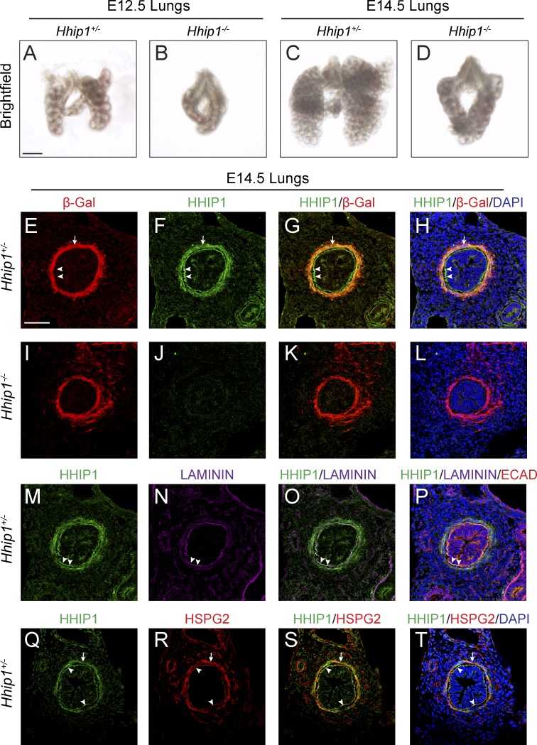
Endogenous HHIP1 protein is secreted and accumulates in the epithelial BM in the embryonic lung. (A–D) Whole-mount images of E12.5 (A and B) and E14.5 (C and D) mouse lungs isolated from Hhip1+/− (A and C) and Hhip1−/− (B and D) embryos. (E–T) Immunofluorescent detection of β-Galactosidase (β-Gal; E, G–I, K, and L), HHIP1 (F–H, J–L, M, O–Q, S, and T), Laminin (N–P), E-Cadherin (ECAD; P), and HSPG2 (R–T) in sections isolated from E14.5 Hhip1+/− (E–H and M–T) and Hhip1−/− (I–L) lungs. (H, L, P, and T) DAPI staining reveals nuclei (blue). Arrows demonstrate overlap between HHIP1 and β-Gal (E–H) or HSPG2 (Q–T) protein expression in the lung mesenchyme. (E–H and M–T) Arrowheads highlight HHIP1 protein staining in the epithelial BM. A commercial HHIP1 antibody (R&D Systems) was used in E–P, whereas a newly developed HHIP1 antibody was used in Q–T. Bars: (A) 500 µm; (E) 50 µm.

