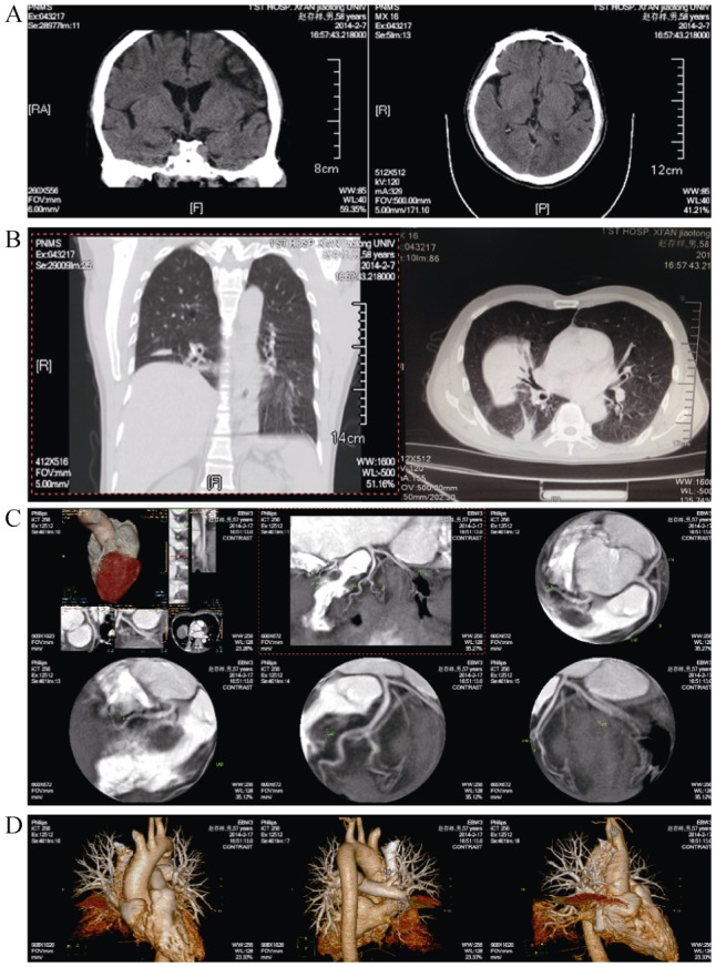
Figure 1. Results of computed tomography and angiography.

(A): Cerebral computed tomography scan showed multiple old lacunar infarcts and brain atrophy; (B): chest computed tomography scan reveals right middle lobe atelectasis, right upper lobe bullae, right lower lobe calcification, right diaphragm elevation and mediastinal lymph node calcification; (C): angiography revealed no significant coronary artery stenosis; (D): angiography revealed no pulmonary embolism and pulmonary arteriovenous fistula.
