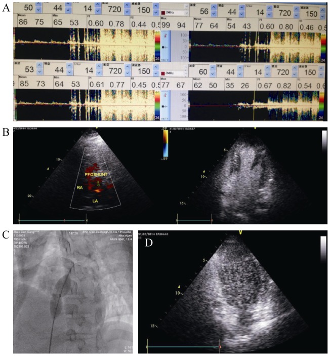
Figure 2. Diagnosis, treatment and subsequent visit of this PFO patient.

(A): TCD foaming test reveals raindrop beaded curtain-like micro-embolism signals lasting approximately 2 min; (B): transthoracic echocardiography and right heart angiography demonstrates left to right shunt in the atrial level and the presence of a patent foramen ovale and numerous micro-bubbles; (C): 24-mm balloon was advanced to determine the size of the PFO and the inflated balloon waist was 15 mm. A closure test was conducted simultaneously and the balloon was set right in the foramen ovale for 30 min under non-oxygen condition; (D): right heart angiography reveals substantial reduction in the amount of micro-bubbles one month after operation. PFO: patent foramen ovale; TCD: transcranial Doppler.
