
Figure 4.
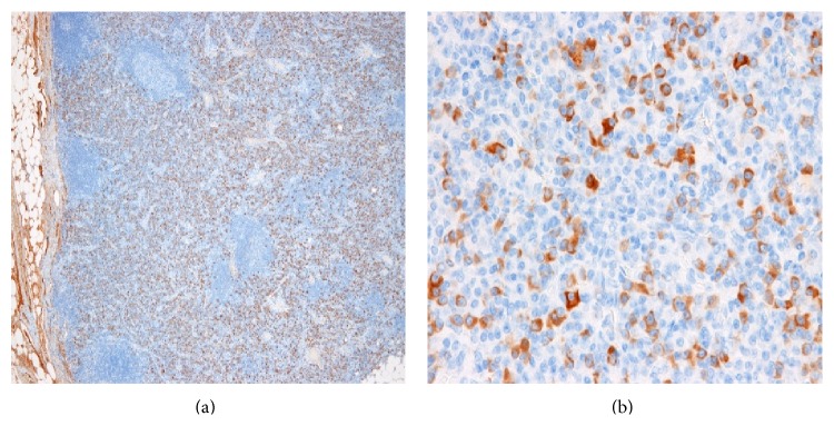
Histopathological findings of the specimen obtained by inguinal lymph node biopsy. IgG4 staining (a, b) revealed IgG4-positive mononuclear cell infiltrates. IgG4/IgG ration was over 90% and more than 300 IgG4-positive plasma cells per high-power field: (a) 4x; (b) 40x.