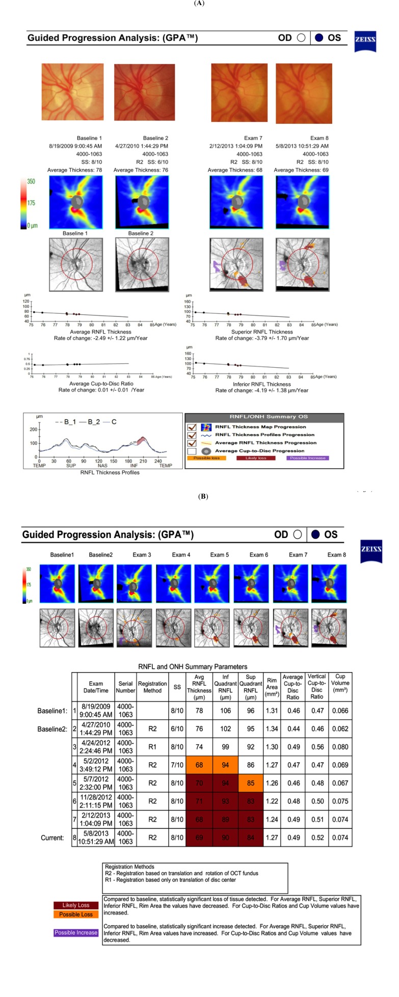
Fig. (2).
Cirrus HD-OCT Guided Progression Analysis (Carl Zeiss Meditec, Dublin, CA, USA) A. Printout showing progressive retinal nerve fiber layer (RNFL) thinning in the superotemporal and inferotemporal regions. Trend lines and rates of change are also provided showing statistically significant slopes of change for RNFL thickness, but not for average cup-to-disk ratio. B. Optic nerve head and RNFL Summary showing worsening of superotemporal and inferotemporal RNFL regions parameters.

