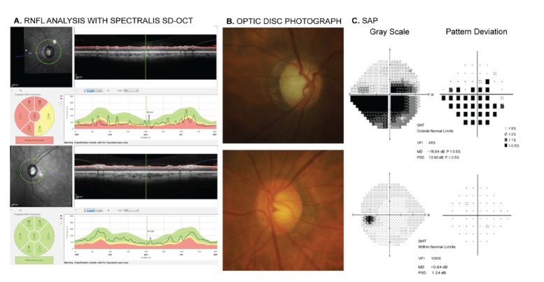
Fig. (1).

Printout of retinal nerve fiber layer (RNFL) analysis obtained with the Spectralis SD-OCT (Heidelberg Engineering, Dossenheim, Germany) in a glaucomatous patient (A). The RNFL evaluation of the right eye shows abnormalities in the superior, temporal and inferior quadrants, which are compatible with the damage seen in the optic disc photograph of this eye (B) and standard automated perimetry (SAP) (C). Evaluation of the left eye showed normal findings on Spectralis SD-OCT, optic disc photograph and SAP.
