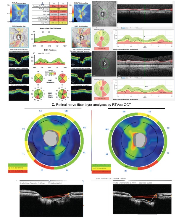
Fig. (3).

Example showing an analysis of the retinal nerve fiber layer (RNFL) of the same subject with three different SD-OCTs: Cirrus-OCT (A), Spectralis SD-OCT (B), and RTVue-OCT (C). The patient had an inferior RNFL defect in both eyes, which was clearly detectable by all three instruments.
