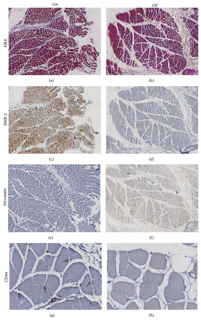
Figure 3.
BMP-2 and satellite stem cells in muscle regenerations. ((a)-(b)) Hematoxylin and eosin sections of muscle biopsies showed a significant increase of fat tissue in OA (a) as compared to OP patients (b) (40x). (c) Image showed numerous BMP-2 positive fibers (40x). Often, in OP patients we did not observed BMP-2 expression (40x) (d). The Immunohistochemistry for myostatin was negative in OA muscle tissue (40x) (e). Muscle biopsies of OP group showed high/moderate expression of myostatin (40x) (f) inversely related to BMP-2 immunostain. Groups of satellite cells CD44 positive were focally dispersed in the tissue (200x) of OA patients (g) higher than that observed in OP patients (200x) (h).

