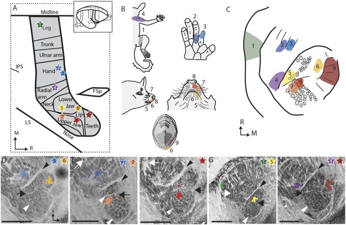
Fig. 4.
Somatotopy of the connections from the ventroposterior nucleus of the thalamus to cortical area 3b. (A) Summary schematic of the nine cortical injection sites (stars) for the tracer CTB in the five hemispheres investigated (four animals). The placement of the injection sites were defined experimentally with 100–300 points in each cortex as well as reference to ref. 42. The stars are numbered and colored to match the rest of the figure. FSp, posterior frontal suclus; IPS, Intraparietal sulcus; LS, lateral sulcus. (B) The multiunit receptive field recorded at the tracer injection sites. (C) A summary schematic of the locations of CTB-stained cell bodies. (D–H) The location of CTB-stained cell bodies (colored dots) mapped onto the adjacent section stained for vesicular glutamate transporter 2 (vGluT2). D–H are all from different hemispheres. Black arrowheads, hand/face border; black arrow, oral fissure border; white arrowhead, barreloid-like vGlut2 clusters; white arrows, hand/foot border. (Scale bars: 1 mm.)

