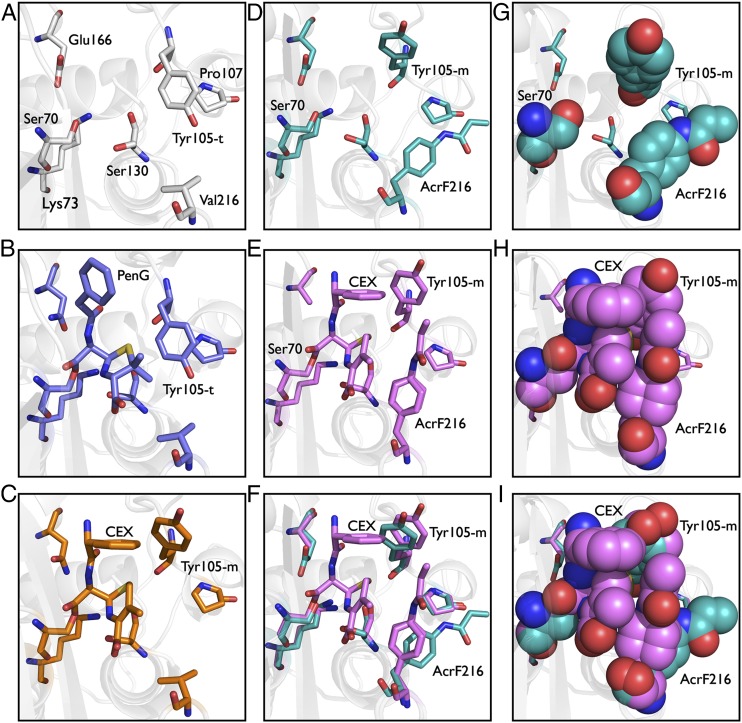
Fig. 3.
Crystal structures of wild-type and Val-216–AcrF mutant β-lactamases. (A) X-ray crystal structure of ligand-free wild-type enzyme (PDB ID code 1BTL). (B) X-ray crystal structure of benzylpenicillin acyl-enzyme intermediate for wild-type enzyme (PDB ID code 1FQG). (C) X-ray crystal structure of cephalexin acyl-enzyme intermediate for wild-type enzyme. (D) X-ray crystal structure of ligand-free Val-216–AcrF mutant enzyme. (E) X-ray crystal structure of cephalexin acyl-enzyme intermediate for the Val-216–AcrF mutant enzyme. (F) Overlay of active-site residues of cephalexin-bound and ligand-free Val-216–AcrF mutant enzymes. (G) X-ray crystal structure of ligand-free mutant enzyme with Val-216–AcrF substitution displayed as cyan spheres. (H) Packing interactions between cephalexin and side chains of mutant enzyme with Val-216–AcrF substitution. (I) Overlay of active site residues as spheres of cephalexin-bound and ligand-free mutant enzymes with Val-216–AcrF substitution.

