Fig. 4.
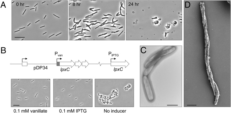
An lpxC-depleted strain twists and lyses. (A) Pvan::lpxC cells were morphologically WT when grown with 0.1 mM vanillate (0 h). Removal of vanillate resulted in a twisted cell morphology by 8 h and ultimately in lysis (24 h). (Scale bar: 10 µm.) (B) lpxC resides in an apparent three-gene operon, and the Pvan::lpxC mutation was complemented by ectopic expression from an IPTG-inducible promoter. (Scale bar: 10 µm.) (C and D) TEM (C) and SEM (D) micrographs show the twisted morphology of the lpxC-depleted strain. (Scale bars: 2 µm.)

