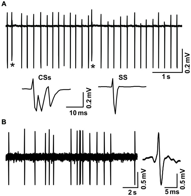
Figure 1.

Cell-attached recordings showing the characterization of cerebellar cortical PCs and MLIs in vivo in mice. (A) Upper, representative cell-attached recording traces showing the spontaneous activity of a cerebellar PC. Lower, enlarged traces of CSs and a simple spike (SS). CSs are indicated by*. (B) Left, representative cell-attached recording traces showing the spontaneous activity of a cerebellar MLI. Right, enlarged traces of spike firing.
