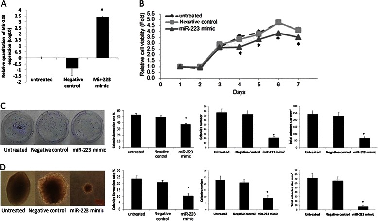
Fig. 2.

Exogenous miR-223 suppresses cell proliferation and colony formation in vitro. a. RT-qPCR result confirmed that the miR-223 expression was significantly higher as compared with untreated and control group after exogenous miR-223 transfection in CNE-2 (*P < 0.05). b. MTT cell viability was assayed one time a day from day 1 to day 7 after the transfection of either miR-223 mimic or the negative control in CNE-2 cells. c. After the transfection with miR-223 mimic or the negative control in CNE-2 cells and the 10 days incubation on plates, the colony formation assay was performed. The colony formation rate, clone number and total clone size were calculated respectively. d. After the transfection with miR-223 mimics or the negative control in CNE-2 cells and the 21 days incubation on soft agar, the colony formation assay was performed. Colonies were counted in 10 randomly chosen microscope fields and the colony formation rate, clone number and total clone size was calculated. We performed at least three independent experiments. The data were showed as means ± SD. * indicates P < 0.05 compared with control group
