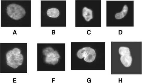
Figure 1.

Patients fibroblasts have been cultured as described in methods. Nuclei have been stained with DAPI and captured using ligth microscope. Examples of abnormal nuclei observed in cells of CMTX1 patients. Normal nuclei (A, B), Abnormal shape (C and D), Polylobbed (E and F). Non disjunction (G and marc).
