Fig. 2.
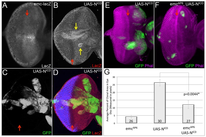
emc is activated by Notch and mediates its growth-promoting activity in the eye. (A-F) Light microscope images of third instar eye-antennal discs containing flp-out overexpression or MARCM clones. Dorsal side is upwards and anterior is towards the right. The red arrows indicate the position of the morphogenetic furrow. All discs were photographed at 10× magnification. (A) Expression pattern of emc-lacZ in a wild-type third instar eye imaginal disc. (B-D) UAS-NICD flp-out clones expressing GFP show increased levels of emc-lacZ in many compartments of the disc. The yellow arrows indicate clones in which emc-lacZ expression is activated in response to Notch signaling. (E,F) MARCM clones expressing GFP induced in a wild-type background. (E) UAS-NICD MARCM clones. (F) emcAP6, UAS-NICD MARCM clones. (G) The average percentage of the eye imaginal disc area occupied by MARCM clones of genotypes listed in E, F and Fig. 1P. Statistical significance was calculated using Student's t-test and equal or unequal variance was determined using a F-test. The difference between UAS-NICD and emcAP6, UAS-NICD clones is statistically significant with a P-value of 0.0044. The number of discs containing clones that were analyzed is listed within the panel. Error bars represent s.d.

