Fig. 6.

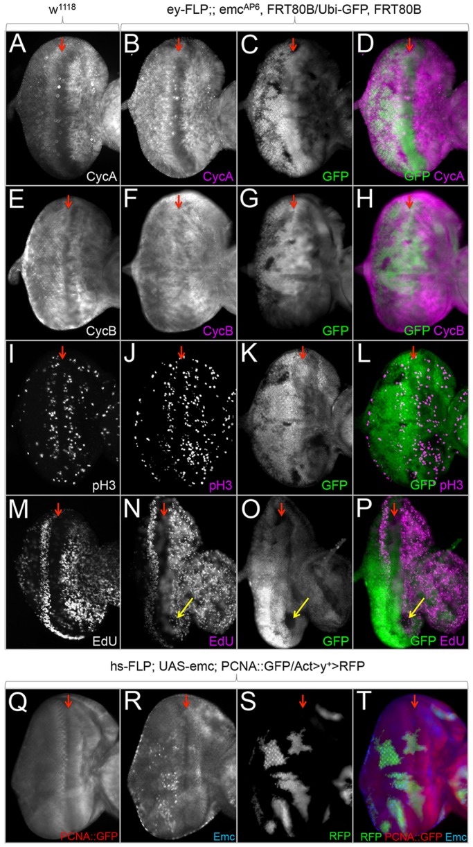
Emc delays entry into S phase in the developing eye. (A-T) Light microscope images of third instar eye-antennal discs containing loss-of-function and flp-out overexpression clones. Dorsal side is upwards and anterior is towards the right. The red arrows indicate the position of the morphogenetic furrow. All discs were photographed at 10× magnification. (A,E,I,M,Q) Normal localization of CycA, CycB and pH3, EdU incorporation, and a readout for E2F in wild-type eye discs. (B-D,F-H,J-L,N-P) Mitotic emcAP6 clones induced continuously throughout eye development using ey-FLP. (B-D,F-H,J-L) emcAP6-null clones (lacking GFP) do not show dramatic alterations in levels of CycA, CycB or pH3 staining. (N-P) emcAP6 clones show decreased number of cells incorporating EdU (yellow arrows) compared with surrounding wild-type tissue. (R-T) Overexpression clones of emc induced with hs-FLP do not show an increase in the PCNA::GFP reporter, indicating that E2F binding to the reporter is not elevated.
