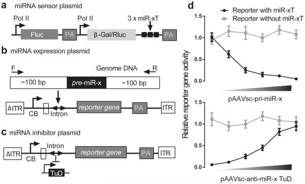
Figure 2.
Validation of miRNA toolkit in HEK293 cells. (A) miRNA sensor plasmid to monitor the miRNA activity. The strategy for construction of AAV plasmids expressing functional pri-miRNA fragments (B) and miRNA inhibitors, TuD RNAs (C). Cross validation of miRNA expression and inhibition plasmids (D). Top at (D) shows increasing amounts of a pri-miRNA producing vector inhibits expression of a miRNA sensor plasmid in HEK293 cells. The bottom shows the de-repression from anti-miR TuDs in a dose response manner when we fix the amount of pri-miR plasmids. ITR, inverted terminal repeat; ΔITR, mutated ITR; CB, chicken β-actin promoter with CMV enhancer; U6, U6 promoter; PA, poly (A); pre-miR, precursor miRNA; Fluc, Firefly luciferase; Rluc, Renilla luciferase; β-Gal, β-galactosidase; 3 × miR-xT, 3 miRNA perfect target sites.

