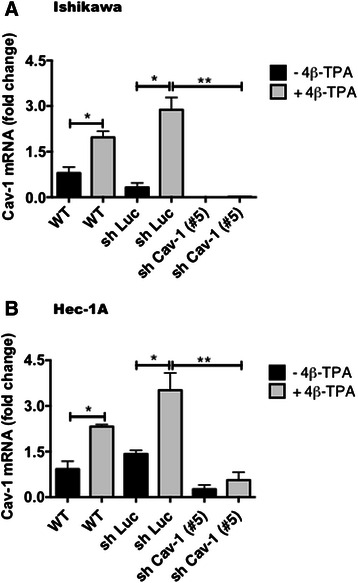
Fig. 2.

CAV1 mRNA levels increased in ECC after exposure to 4β-TPA. Ishikawa and Hec-1A cells were transduced with CAV1 shRNA (shRNA Cav-1-(#5)) or shRNA for Luciferase (shLuc), as a control. Stably transduced cells expressing the corresponding construct were obtained by selection in medium with puromycin. Wild type or transduced Ishikawa (a) and Hec-1A (b) cells were seeded in 6-cm dishes for 24 h in complete medium and then cultured in medium without serum for 24 h or 48 h, respectively, prior to 100 nM 4β-TPA stimulation for 24 h. CAV1 mRNA levels were assessed by real time RT-PCR analysis. β-actin was used as an internal control. Values obtained by analysis of three independent experiments are shown for CAV1 mRNA following standardization to β-actin (mean ± SEM). Data were analyzed using the unparied t-test. Statistically significant differences compared with the controls are indicated (*, p < 0.05, **,p < 0.01). Note that levels detected for the wild type (WT) cells without 4β-TPA were assigned the reference value 1
