Fig. 6.

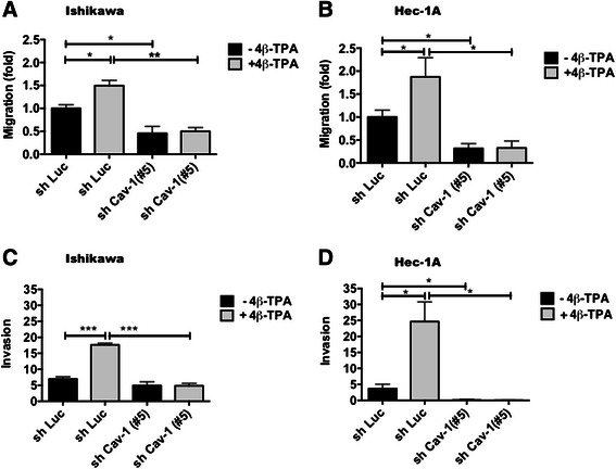
CAV1 expression in ECC augmented transmigration and invasion. To evaluate the role of the up-regulation of CAV1 mediated by 4β-TPA in transmigration 6 × 10⁵ shLuc and shCav-1(#5) Ishikawa or Hec-1A cells were seeded in 6 cm plates in complete medium for 24 h prior to serum withdrawal for an additional 24 or 48 h of culture, respectively. After 24 h of 100 nM 4β-TPA treatment 2 × 10⁵ shLuc and shCav-1(#5) Ishikawa (a) or Hec-1A (b) cells were seeded in Boyden Chambers coated on the lower side with fibronectin (2 μg/ml) and allowed to migrate in the absence of serum for 7.5 h. The cells that migrated through the pores and bound to the lower fibronectin-coated surface were stained and counted. Values obtained were normalized to the shLuc cells without treatment. The number of transmigrated shLuc cells observed in panels a and b were 48 ± 9 and 94 ± 26, respectively. Averages from three independent experiments are shown (mean ± SEM). Statistically significant differences compared with the corresponding control group are indicated (*, p < 0.05). To evaluate the role of the up-regulation of CAV1 mediated by 4β-TPA in invasion, shLuc and shCav-1(#5) Ishikawa and Hec-1A cells were seeded in 6 cm plates in complete medium 24 h prior serum withdrawal for an additional 24 or 48 h of culture, respectively. After 24 h of 100 nM 4β-TPA treatment 2 × 10⁵ shLuc and shCav-1(#5) Ishikawa (c) and Hec-1A (d) cells were seeded over matrigel covered porous inserts, with or without 100 nM 4β-TPA for 24 h. The number of cells that invaded the matrigel and migrated through the pores were determined after immunostaining for cytokeratin. Averages from three independent experiments are shown (mean ± SEM). Statistically significant differences compared with the corresponding control group are indicated (*, p < 0.05, **, p < 0.01, ***, p < 0.001)
