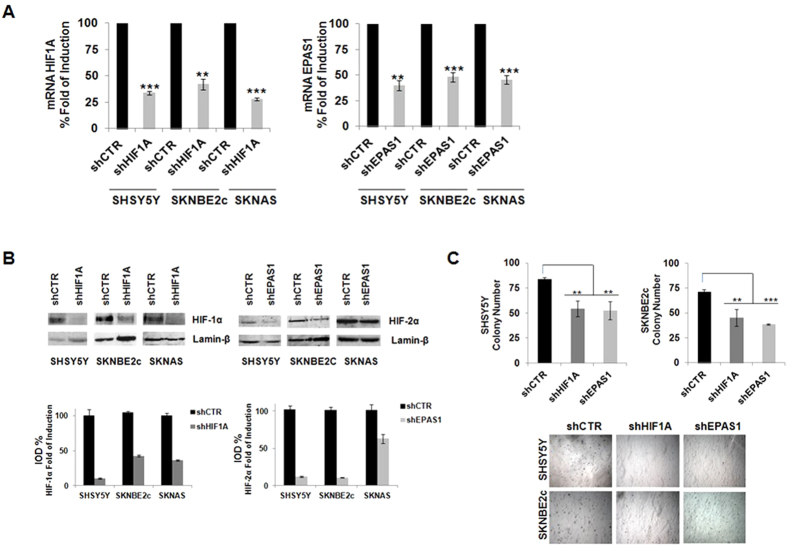
Figure 4. HIF1A and EPAS1 silencing in NBL cells and the effects on cell growth in the colony formation assay.
(A) Efficiency of gene silencing mediated by lentiviral delivery of hairpin RNAs directed against HIF1A and EPAS1 (shHIF1A, shEPAS1, respectively) in the three NBL cell lines (SHSY5Y, SKNBE2c, SKNAS) was assessed using RT-PCR. Data are means of three experiments and are represented as percentages with respect to the NBL shCTR cells, which were infected by lentivirus-mediated delivery of non-silencing hairpin RNA. (B) Western blotting of nuclear extracts from HIF1A and EPAS1 silenced cells showing the decreases in the HIF-1α and HIF-2α proteins. The bands were quantified by densitometry. The bar graphs show the integral optical density (IOD) for each band, normalized with respect to lamin-β expression. (C) HIF1A and EPAS1 silencing in SHSY5Y and SKNBE2c cells affects cell growth in the soft agar colony formation assay. The graft bars show decreased colony numbers in the NBL shHIF1A/shEPAS1 cells compared to the NBL shCTR cells. The SKNAS cells are not shown (** P ≤ 0.01; *** P ≤ 0.001).

