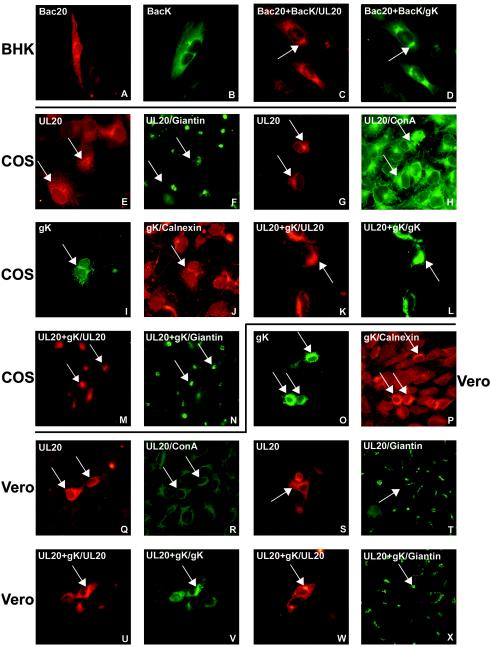
FIG.1.
Intracellular localization of UL20p and gK detected by immunofluorescence. (A to D) BHK cells transduced with baculovirus-UL20 (Bac20), baculovirus-gK (BacK), or both and stained with PAb to UL20p (A and C) or PAb to myc (B and D). (E to X) Two paired images, one green and one red, are shown for the same photographic field, and the paired panels are E and F, G and H, I and J, K and L, M and N, O and P, Q and R, S and T, U and V, and W and X. (E to H) COS cells transfected with pUL20-pcDNA and stained with PAb to UL20p with (F) and without (E) Ab to giantin and with (H) and without (G) ConA-FITC. (I and J) COS cells transfected with pgK and stained with anti-myc Ab with (J) or without (I) Ab to calnexin. (K to N) COS cells cotransfected with pUL20-pcDNA and pgK and stained with Ab to UL20p (K and M), myc (L), or giantin (N). (O and P) Vero cells transfected with pgK and stained with Abs to myc (O) or calnexin (P). (Q to T) Vero cells transfected with pUL20-pcDNA and stained with Ab to UL20p (Q and S), anti-giantin (T), or ConA-FITC (R). (U to X) Vero cells cotransfected with pUL20-pcDNA and pgK and stained with Ab to UL20p (U and W), myc (V), or giantin (X). Cells were transfected with 250 ng of each plasmid. Cells transfected with a single plasmid received an additional 250 ng of pMTS. The protein indicated after the slash identifies the stained protein in double immunofluorescence.

